
Case Report: Managing a Rare Parapharyngeal Ganglioneuroma in a Young Girl
Sometimes in medicine, the most interesting stories don’t start with symptoms, but with coincidence. This case is no exception. It all started with a simple visit to the emergency room after a head injury. In a sixteen-year-old girl, a routine CT scan unexpectedly revealed a large tumor hidden deep in her neck.
The case report was published in November under the title “Incidental parapharyngeal ganglioneuroma: A case report on surgical management.” The authors are Michael G. Hahn, Courtney J. Hunter, and André M. Wineland.
Ganglioneuroma, a rare and benign tumor
The tumor was identified as a ganglioneuroma, a rare and benign tumor arising from the sympathetic nervous system. Ganglioneuromas account for only 6.4 percent of peripheral neural crest tumors and are most commonly found in the abdomen, pelvis, or chest. Their occurrence in the parapharyngeal space, a region of the neck adjacent to the pharynx and major blood vessels, is very rare. Fewer than 40 cases have been reported in the medical literature over the past four decades, and this rarity has limited both clinical experience and the development of clear management guidelines.
In this patient, a non-contrast CT scan of the head demonstrated a large mass in the right retropharyngeal space. A subsequent contrast-enhanced CT scan of the neck confirmed a lesion measuring 5.2 × 3.9 × 2.0 centimeters. Notably, the patient had minimal symptoms. She reported occasional difficulty swallowing solid food. “Due to their slow-growing nature, parapharyngeal ganglioneuromas often remain asymptomatic until reaching a significant size with resulting mass effect,” the scientists wrote.
Magnetic resonance imaging further clarified the extent and location of the lesion. Based on the imaging findings and clinical context, a surgical excisional biopsy was recommended to establish a definitive diagnosis. The mass was removed using a transoral (through the mouth), endoscope-assisted approach. The procedure was completed without complications. Histopathological examination confirmed the expected diagnosis: a mature, benign ganglioneuroma.
Three months later
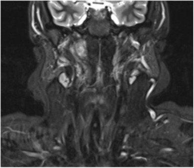
A follow-up MRI performed three months after surgery showed a smaller residual portion of the tumor located in the right parapharyngeal and retropharyngeal space, adjacent to the internal carotid artery, one of the major vessels supplying blood to the brain. In cases involving benign tumors, surgeons may prioritize safety over total removal. If a tumor is tightly adhered to a critical structure, like this artery, it is often safer to leave a small fragment behind rather than risk life-threatening injury. Because the tumor is non-cancerous and grows very slowly, the remaining piece can be safely monitored. Although the residual tumor remained in contact with the artery, blood flow was preserved. Clinicians elected to pursue careful long-term surveillance using serial imaging.
“Despite the rarity of this entity, the case highlights the importance of a comprehensive diagnostic workup, including imaging studies and histopathological examination, to accurately differentiate it from other parapharyngeal space lesions,” they concluded in the report.
Image: The MRI scan shows the portion of the tumor that remained after surgery. Hahn M.G., Hunter C.J., Wineland A.M. Incidental parapharyngeal ganglioneuroma: A case report on surgical management, Arkansas Children’s Hospital & University of Arkansas for Medical Sciences, 2025.