
Protecting Boys’ Fertility: Emily Delgouffe at VUB
Earlier this year, a historic medical breakthrough was achieved when surgeons successfully performed the first transplant of immature testicular tissue, which was preserved for 16 years. Every year, an estimated 400,000 children and adolescents aged 0 to 19 develop cancer, making such medical achievements particularly significant. Children who have undergone treatments such as chemotherapy or radiation in childhood can lose their fertility due to damage to the testicles or ovaries, and this loss of fertility can be temporary or permanent. The procedure was performed on a patient who had received chemotherapy during childhood to restore his fertility, under the guidance of Professor Ellen Goossens, Emilie Delgouffe’s main supervisor.
Meet Emily Delegouffe
In 2002, Emily Delgouffe was only five years old; while she was still discovering the world through play, one of the first clinical programs in the world for freezing immature testicular tissue was launched at BITE (VUB). Emily could not have imagined that, years later, she would walk the halls of Vrije Universiteit Brussel (VUB), earn her doctorate there, and dedicate her work to the very field that had just begun that year.
When asked if the clinical program initiated in 2002 prepared the ground for the generations that came and those yet to come, Emily says that, although she was only five at the time, looking back, it was a big step forward for the field and laid the foundation for everything they do today. “By the time I started my research, the infrastructure, clinical protocols, and biobanking systems were already in place. Thanks to that early start, we now have a unique patient population for long-term follow-up, which allows us to closely link clinical care with translational research. It has not only paved the way for our generation of researchers but also for future patients who may one day benefit from fertility restoration techniques. We’re now actively working on developing and optimizing those restoration methods-something that would not have been possible without that early clinical vision,” she says proudly.
On the question of what a typical week in the laboratory looks like for her when she is at her busiest, she said that her most demanding weeks revolve around grant deadlines. “Writing proposals, coordinating with collaborators, and preparing supporting data take up most of my time. I’m also finalizing publications and preparing for a new research project, which means I’ll soon be back at the bench. In parallel, I’m helping PhD students with their work and getting ready to supervise new Bachelor’s and Master’s students in the upcoming academic year,” she added.
“We have easy access to shared infrastructure and core facilities, which allows us to make optimal use of advanced technologies without unnecessary duplication. This flexibility and openness make it easier to set up interdisciplinary projects and respond quickly to new research opportunities.”
What does VUB do well when it comes to this research, and what is the university’s greatest strength in this area?
Emily Delgouffe: One of VUB’s key strengths is the close collaboration between hospital and research teams. This strong link between clinical care and fundamental science allows us to translate patient needs directly into research questions-and vice versa. It also enables us to follow patients longitudinally and integrate clinical data with experimental findings. Another advantage is the collaborative mindset across labs and departments. We have easy access to shared infrastructure and core facilities, which allows us to make optimal use of advanced technologies without unnecessary duplication. This flexibility and openness make it easier to set up interdisciplinary projects and respond quickly to new research opportunities.
“What fascinates me most is how testicular cells, even without a printed support, naturally “know” how to find each other and self-organize into organoids. This remarkable self-assembly highlights the innate biological programming that enables cells to recreate complex tissue structures, which holds exciting potential for tissue engineering.”
In the first year of your master’s degree, you joined the testicular biology research group for an internship on generating supports for in vitro spermatogenesis in humans using 3D bioprinting. How accurate is today’s technology in preserving living cells during printing, and what fascinated you the most?
Emily Delgouffe: Previous studies in our lab, performed in mice, attempted to bioprint interstitial cells directly into scaffolds, but these cells could no longer be detected after culture. This suggested that the printing process, or the low cell numbers, negatively affected cell survival or retention. Since further increasing the cell concentration in the bioink was not possible in human research due to limited tissue availability, we moved away from printing cells altogether. Therefore, during my internship, we printed only the scaffold using an alginate-based bioink and then seeded human interstitial cells onto it. These cells formed extracellular matrix-producing aggregates, showing they could interact with the scaffold. The next step was to use a sacrificial bioink such as PLURONICS, which could be degraded by temperature changes to remove the scaffold after interstitial cell reorganization, allowing the seeding of spermatogonial stem cells in the resulting compartments.
Unfortunately, this approach was not feasible because the high water content of the agarose support caused the PLURONICS bioink to slip during printing. Because of these technical challenges, high costs, and the complexity of bioprinting, we shifted our focus to an alternative method using agarose microwells. This approach does not require bioprinting but still allows spatial control and 3D organization of testicular cells. It has already yielded promising results in mouse testicular organoids, including spermatogenesis, testis-specific architecture, improved germ cell survival, and testosterone production. What fascinates me most is how testicular cells, even without a printed support, naturally “know” how to find each other and self-organize into organoids. This remarkable self-assembly highlights the innate biological programming that enables cells to recreate complex tissue structures, which holds exciting potential for tissue engineering.
“What surprised me most was how unpredictable some outcomes could be. Some patients who had received very intensive treatment still showed active sperm production in adulthood. Even more striking was that two patients with the same diagnosis and the same treatment could have completely different outcomes. One might be producing normal sperm just a few years after treatment, while another remains infertile more than ten years later. This shows how complex and individual reproductive recovery can be.”
During your PhD, you examined the pubertal development and fertility of boys who underwent a testicular tissue biopsy in the context of fertility preservation. Why did you choose this topic? What particularly surprised you, and what was the biggest issue?
Emily Delgouffe: This topic felt like a natural choice. Our hospital was the first in the world to offer immature testicular tissue banking as a clinical fertility preservation strategy, starting in 2002. Thanks to that early start, we now have a unique group of patients we can follow up with many years after their treatment. This long-term perspective is essential because although freezing testicular tissue offers hope for future fertility, the technique is still experimental. The procedure involves surgery, and we do not yet fully understand its long-term effects. In animal studies, transplanting frozen–thawed testicular tissue has already led to the birth of healthy offspring. In humans, however, we are still waiting to see whether this approach can truly restore fertility. That is why it is so important to understand which patients are most likely to benefit from this technique and to ensure that we offer it to the right individuals.
What surprised me most was how unpredictable some outcomes could be. Some patients who had received very intensive treatment still showed active sperm production in adulthood. Even more striking was that two patients with the same diagnosis and the same treatment could have completely different outcomes. One might be producing normal sperm just a few years after treatment, while another remains infertile more than ten years later. This shows how complex and individual reproductive recovery can be.
One of the biggest challenges in this research is the limited number of patients available for long-term follow-up. In addition, the patient population is very heterogeneous, with differences in diagnosis, age at treatment, and treatment protocols. This makes it difficult to determine which specific factors are responsible for the observed effects on fertility. As a result, drawing clear conclusions remains challenging. This highlights the importance of collaboration between centers so that data can be pooled and compared across larger and more homogeneous groups. Only through such efforts can we refine our understanding and improve patient selection for testicular tissue banking.
“Continued research, better risk prediction, and long-term follow-up are crucial to improve outcomes and ensure that fertility preservation is offered to the right patients at the right time.”
As noted in the study “Testicular Tissue Banking for Fertility Preservation in Young Boys: Which Patients Should Be Included,” a major complication is the damage to the germ cells and testicular somatic cells caused by the gonadotoxic properties of chemo- and/or radiotherapy. In other words, cancer drugs and radiation can destroy cells in the testes, causing some men to have fertility problems, sometimes only temporarily, and sometimes permanently. What are the possible solutions? Is there a way to improve current therapies, and to what extent is that realistically achievable?
Emily Delgouffe: Reducing the impact of cancer treatment on fertility is an important goal, especially in children, but it remains a complex challenge. Survival always comes first, so reducing chemotherapy doses or switching to less harmful agents is only possible in selected cases. Radiotherapy techniques have become more precise, and approaches like proton therapy can reduce exposure to surrounding healthy tissue. In children, this may help limit testicular damage, but the testes are extremely sensitive to radiation, and even low doses can impair fertility.
Another area of interest is the development of protective agents that could avoid damage to testicular cells during treatment. However, these drugs are still in the experimental phase and not yet available in clinical practice. Realistically, completely avoiding gonadotoxic effects is unlikely in the near future. That is why fertility preservation remains essential. For prepubertal boys, testicular tissue banking is currently the only available option. Continued research, better risk prediction, and long-term follow-up are crucial to improve outcomes and ensure that fertility preservation is offered to the right patients at the right time.
“To advance the field, we need robust long-term data on safety and efficacy, as well as standardized inclusion criteria and improved risk stratification. Currently, patient selection varies widely between centers due to a limited understanding of which treatments truly pose a high risk of infertility. While patient advisory committees are often highly enthusiastic and supportive of this research, that enthusiasm unfortunately does not always translate into concrete funding. Sustained investment and international, multicenter collaboration remain essential to move testicular tissue banking from an experimental approach to a clinically established practice.”
In your doctoral research, “Effects of gonadotoxic treatments on spermatogonial stem cells and their niche,” it is mentioned that “immature testicular tissue banking is classified as an invasive and experimental procedure whose long-term effects and clinical applications have yet to be thoroughly investigated.” What changes are necessary, and is there currently sufficient investment in this field of research?
Emily Delgouffe: Testicular tissue banking in young boys remains experimental until we can demonstrate that fertility restoration methods are truly effective. We are not there yet, but the field is moving in the right direction. Recently, previously frozen testicular tissue was autologously reimplanted in a patient at our own center (Het Academisch Ziekenhuis Brussel (H.U.B.), and spermatogonial stem cells were re-transplanted in another patient in the United States, both using material cryopreserved years earlier. These are the first clinical applications of this technique in humans, though outcomes are still pending.
To advance the field, we need robust long-term data on safety and efficacy, as well as standardized inclusion criteria and improved risk stratification. Currently, patient selection varies widely between centers due to a limited understanding of which treatments truly pose a high risk of infertility. While patient advisory committees are often highly enthusiastic and supportive of this research, that enthusiasm unfortunately does not always translate into concrete funding. Sustained investment and international, multicenter collaboration remain essential to move testicular tissue banking from an experimental approach to a clinically established practice.
“International collaboration is key to overcoming the limitations of small and diverse patient cohorts. Existing studies often lack generalizability due to limited sample sizes and variation in diagnoses and treatments. To address this, the ORCHID-NET consortium was launched in 2022. It brings together male fertility preservation experts to develop evidence-based guidelines and coordinate long-term follow-up.”
One of the main challenges in studies like this is the small number of patients, along with the variability in diagnoses and treatment types. Could international collaborations help overcome these limitations? Are there existing databases for this type of research, and is the available data sufficient?
Emily Delgouffe: Yes, international collaboration is key to overcoming the limitations of small and diverse patient cohorts. Existing studies often lack generalizability due to limited sample sizes and variation in diagnoses and treatments. To address this, the ORCHID-NET consortium was launched in 2022. It brings together male fertility preservation experts to develop evidence-based guidelines and coordinate long-term follow-up. One of its core goals is to create a comprehensive international patient registry. Such registries will help standardize data collection, confirm treatment-related gonadotoxicity, and refine inclusion criteria for testicular tissue banking. While some local datasets exist, the available data remain fragmented and insufficient.
“Personally, I would like to see greater public awareness about fertility preservation and its importance, more consistent follow-up of survivors into adulthood, and improved integration of fertility care into survivorship programs. Using research findings to improve patient care is a challenge due to the complexity of the issue, but with better collaboration and long-term data collection, it is achievable and crucial for improving quality of life.”
In Europe, about 35,000 new cases of childhood and adolescent cancer are reported each year, leading to nearly half a million cancer survivors between 2020 and 2025. This means that an increasing number of adults will have to deal with the long-term effects of cancer treatments. Are we paying enough attention to this topic in Europe? Are there enough studies and results that could help in the future? What would you personally like to see change? Is it a challenge to actually use research findings to improve people’s lives?
Emily Delgouffe: At our hospital, informing patients and their parents about the potential impact of cancer treatment on fertility is routine. Across Europe, fertility preservation is increasingly being integrated into oncology programs, improving awareness and care within specialized centers. However, it is still not standard practice in all healthcare settings. The general public is much more aware of immediate side effects, such as hair loss or fatigue, while fertility loss often remains unknown and can come as a shock to patients and families. Research on long-term fertility outcomes after childhood cancer is growing, but challenges remain. Patient groups are small and diverse, treatment protocols vary widely, and long-term follow-up data are often lacking. This makes it difficult to translate research findings into standardized care that benefits all patients.
Personally, I would like to see greater public awareness about fertility preservation and its importance, more consistent follow-up of survivors into adulthood, and improved integration of fertility care into survivorship programs. Using research findings to improve patient care is a challenge due to the complexity of the issue, but with better collaboration and long-term data collection, it is achievable and crucial for improving quality of life.
What is your favorite research method, and why? Do you think enough is being done to develop new research methods in this field?
Emily Delgouffe: I’m very interested in transcriptomic techniques. So far, I’ve only briefly worked with bulk RNA sequencing, which already provides valuable insights into gene expression in testicular tissue. But I’m especially excited to explore more advanced methods like single-cell and spatial transcriptomics during my postdoc. These techniques allow for much more precise analysis of complex tissues, which is crucial in reproductive research where cell-cell interactions and spatial context matter. Sequencing technologies are evolving rapidly, and I think the field is moving in the right direction. That being said, many of these newer methods are still expensive, technically demanding, or not yet optimized for small or rare human samples. I would love to see more efforts to make these tools more accessible and better adapted to clinical and translational research.
“In Belgium, the costs for the biopsy are covered by the national health insurance, and storage is funded by the hospital (UZ Brussel), making it accessible for patients within clinical and research settings.”
Is the preservation of testicular tissue an expensive procedure? How accessible is it for patients in terms of cost and availability?
Emily Delgouffe: Testicular tissue banking is a relatively expensive procedure due to the surgical biopsy, cryopreservation, and long-term storage. However, in Belgium, the costs for the biopsy are covered by the national health insurance, and storage is funded by the hospital (UZ Brussel), making it accessible for patients within clinical and research settings. In other countries, coverage varies. Some rely on public healthcare systems or research funding, while in others, patients may need to cover part of the costs themselves. Overall, accessibility depends on local infrastructure, funding models, and whether the procedure is offered as part of a clinical or experimental program.
“A major missing link is understanding what human testicular cells need in vitro to fully support spermatogenesis.”
When discussing the development and function of the testes, what key aspects or ‘missing links’ still need to be understood?
Emily Delgouffe: A major missing link is understanding what human testicular cells need in vitro to fully support spermatogenesis. In the lab, we can keep testicular cells alive and even mimic the early steps, but full differentiation into mature sperm cells remains a challenge. We don’t yet know exactly which signals or support the cells are missing outside the body—whether it’s specific hormones, structural cues, or interactions between the right somatic cells at the right time. Figuring this out is crucial to eventually achieve human in vitro spermatogenesis, which could offer fertility options for boys who had cancer treatment before puberty.
“Future research should aim to transition testicular tissue banking from an experimental procedure to a clinically validated strategy for fertility restoration.”
On the question of what direction further research should take, Emily stated that future research should aim to transition testicular tissue banking from an experimental procedure to a clinically validated strategy for fertility restoration. According to her, the key areas of focus include the optimization and clinical validation of restoration techniques such as autologous tissue (tissue that comes from the patient’s own body) and spermatogonial stem cell transplantation, and, in the longer term, in vitro spermatogenesis.
“Simultaneously, there is a pressing need for long-term follow-up studies to evaluate both endocrine function and fertility outcomes following tissue preservation and childhood cancer treatments. Establishing large, standardized datasets through international collaboration and data sharing will be critical for developing evidence-based inclusion criteria for testicular tissue banking. In parallel, research must address the reproductive risks associated with emerging cancer therapies such as immunotherapy, which is increasingly used in pediatric oncology. Their potential impact on fertility remains poorly understood and warrants thorough investigation. Ultimately, integrating long-term patient monitoring with preclinical drug testing will enhance our ability to identify which patients are genuinely at risk of infertility. This will enable more targeted and meaningful application of testicular tissue banking, ensuring that the intervention is only offered to those who will benefit most,” she concluded.
New Recommendations and Guidelines for Fertility Preservation in Boys and Adolescents
In a 2024 study with her colleagues, Professor Gossens provided an overview of how fertility preservation in boys and adolescents is practiced around the world. As they report, since 2002, 16 centers have preserved testicular tissue from 3,118 boys under the age of 18. The approach varies from center to center in terms of patient selection, storage, and follow-up. Globally, over 3,000 boys have had tissue preserved; however, it is important to note that not all centers were included, so the actual number is likely higher.
In August 2025, Professor Gossens contributed to “ESHRE good practice recommendations on fertility preservation involving testicular tissue cryopreservation in children receiving gonadotoxic therapy, ” a study that further enriched the field. The question they addressed was how fertility preservation in male children and adolescents receiving gonadotoxic therapies (therapies that can cause temporary or permanent damage to the ovaries or testes) should be managed. Forty-four recommendations were formulated to guide all aspects of fertility preservation in prepubertal boys and adolescents. As the authors emphasize, these guidelines offer valuable support for healthcare professionals, aiming to promote knowledge among clinicians while enabling patients to make informed decisions based on realistic expectations.
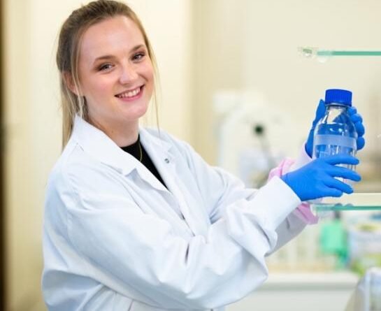
Image: PhD Emiliy Delgouffe/VUB
With Emilie Delgouffe’s scientific journey as our starting point, we began with the publication of the first of 21 articles. These publications were produced as part of the Maria Leptin EMBO Fellowship, which allowed us to spend two months exploring the world of science at VUB in Brussels. Importantly, all articles were the result of our own choice of topics and in accordance with our interests.