
Bladder Melanosis in a Child: The Second Reported Case
Bladder melanosis is a rare condition in which the pigment melanin accumulates in the bladder. By itself, it is usually not dangerous and does not indicate cancer. The dark spots can look similar to cancer, so a biopsy is needed to rule it out. It often occurs with chronic bladder irritation or inflammation. Because of the small number of cases, the long-term consequences are still unknown.
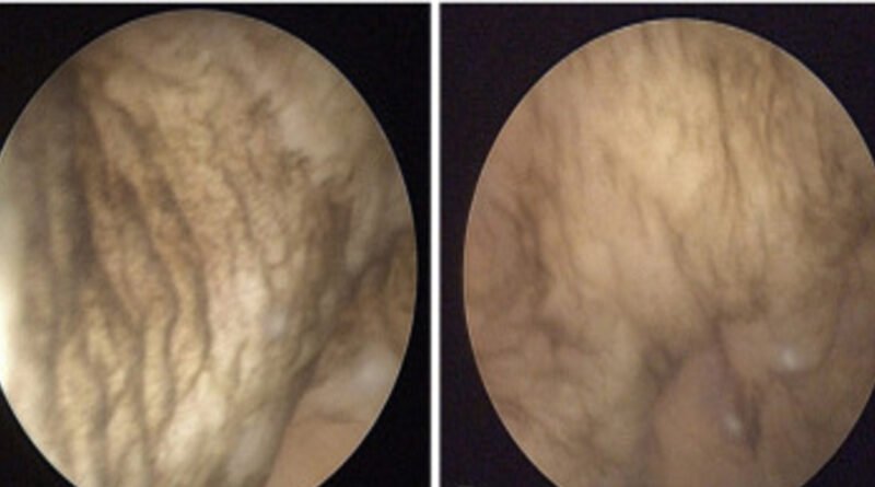
Fewer than 40 cases have been reported, and only one previously occurred in children. Recently, the scientists presented a second case in a 13-year-old boy. Cystoscopy showed brown pigmentation, and biopsy confirmed melanosis with chronic inflammation, without signs of malignancy. The lesion resolved spontaneously, without the need for treatment.
The case report is titled ‘Paediatric bladder melanosis: a rare case report.’ The authors are Suzannah Woodhouse, Alexander Cho, Athanasios Tyraskis, Sharmila Sanchez, and Thivya Sekar.
As the authors explained, bladder melanosis was first described by Alroy et al. in 1986. They note that, with fewer than 40 documented cases worldwide, the underlying etiology (cause of the disease) and potential long-term clinical implications remain poorly understood.
About the case
The report states that a 7-year-old boy presented with intermittent urinary bleeding. An ultrasound showed a thickened and irregularly shaped bladder, but the kidneys were normal. Cystoscopy revealed a type of blockage in the urethra, which was removed. After three months, the bladder was less thickened but still larger than normal.
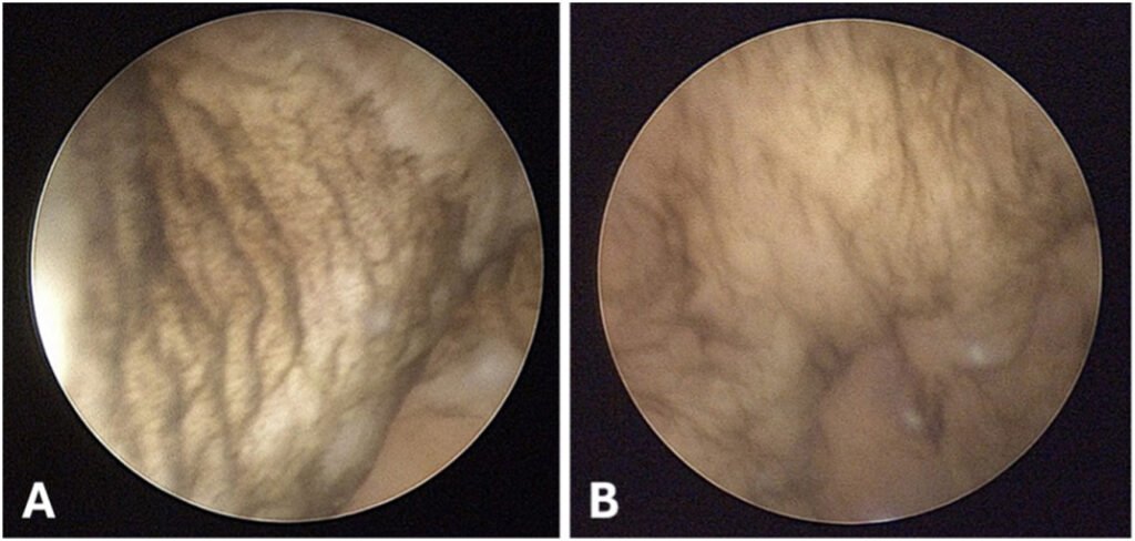
The boy was followed up regularly for the next five years. At age 13, an ultrasound showed mildly dilated ureters and small changes in the bladder due to long-term high bladder pressure. Cystoscopy revealed dark spots in the bladder, and a biopsy showed the presence of melanin and mild inflammation, but no cancer. The patient recovered well, and a follow-up cystoscopy four months later showed that the dark spots had completely disappeared.
The only case in the paediatric reported literature that shows macroscopic resolution
“Our case is the only case in the paediatric reported literature that shows macroscopic resolution of the melanosis on subsequent cystoscopy and microscopic resolution on histology,” they wrote.
What is important for both physicians and the general public to know is that there is currently no standardized approach to investigating, treating, or following up on this condition. As a result, treatment decisions are left to the medical team. In the conclusion, they emphasize the essential role of histopathological analysis in distinguishing benign from malignant bladder lesions. It also highlights that ongoing case reports and research are crucial for improving the understanding of the clinical significance of this rare condition.
Image credit: The study