

Two Cancers in One Kidney: A Rare Case That Challenged Diagnosis
When doctors discover a tumor, the first step is usually to determine its size, location, and whether it has spread. However, sometimes one tumor is not just one tumor.
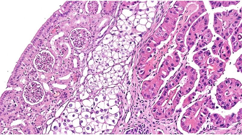
In a rare case involving a 62-year-old man, pathologists found two different types of cancer in the same kidney after surgery. One was clear cell renal cell carcinoma, the most common subtype of kidney cancer, while the other was type 2 papillary renal cell carcinoma. Both tumor components were located in the same region of the kidney, but they were clearly separated.
The case was reported by Kays Chaker, Ayda Khadhar, Boutheina Mosbahi, Afif Limem, Adel Ammous, and Yassine Nouira in “A Collision of Two Primary Renal Neoplasms: A Rare Case Report.”
Such neoplasms are known as renal collision tumors, tumors in which two histologically different neoplasms occur within the same organ without a clear transitional area between them.
Two Cancers in One Kidney
The patient, a 62-year-old man and smoker with diabetes and hypertension, presented with blood in his urine that had persisted for approximately six months. He also had right flank pain, and a physical examination revealed a palpable mass.
A blood test showed a hemoglobin level of 8.6 g/dL, indicating significant anemia. Computed tomography revealed a heterogeneous mass in the upper pole of the right kidney, measuring approximately 6.5 centimeters. However, the scan did not allow doctors to reliably determine that the mass contained two different types of cancer. It appeared to be one large, heterogeneous renal neoplasm. The patient underwent a radical right nephrectomy, meaning surgical removal of the entire kidney.
Only then was the unusual nature of the tumor revealed. Microscopic examination showed two clearly separated components.
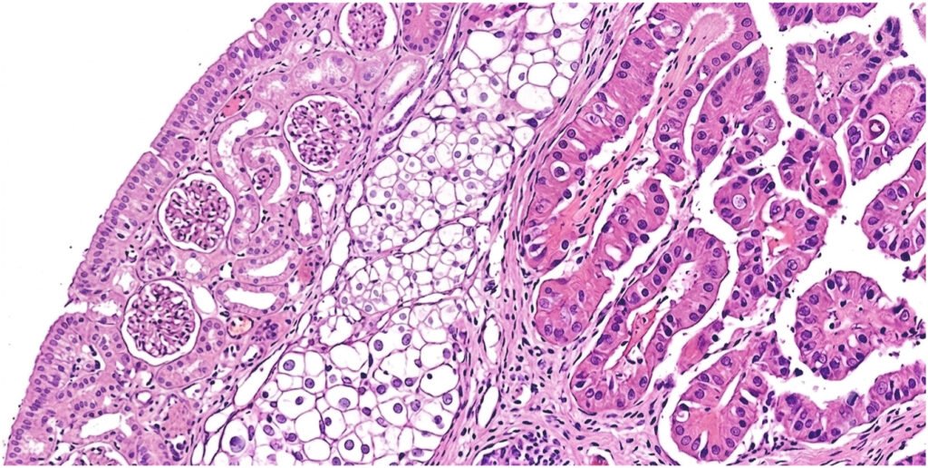
The first was clear cell renal cell carcinoma, measuring six centimeters. It was confined to the kidney, with no invasion through the renal capsule or into the surrounding fatty tissue. The second component was type 2 papillary renal cell carcinoma, measuring seven centimeters. Unlike the first tumor, this one had invaded the fat surrounding the kidney.
The pathological findings were particularly important: there was a clear boundary between the two tumor components, with no area in which one type of cell gradually transitioned into the other. This characteristic is crucial for the diagnosis of a collision tumor.
How Do Two Tumors End Up Together?
Medicine still does not have a definitive answer to this question.
For this reason, collision tumors represent an intriguing problem not only for pathologists but also for researchers trying to understand how different forms of cancer develop. The challenge begins with radiological imaging. CT and magnetic resonance imaging can show the size and structure of a tumor, as well as how different parts of the tumor behave after contrast administration. However, these characteristics are usually not sufficient to reliably distinguish between different histological subtypes. It is also important to note that heterogeneity does not automatically mean that two different types of cancer are present within the tumor.
This is why the definitive diagnosis of a collision tumor often comes only after surgical removal and detailed pathological examination.
A Rarity That Raises a Broader Question
Renal collision tumors are rare enough that their behavior still cannot be predicted with precision. Their rarity is precisely what makes them difficult to study. Most available data come from individual case reports rather than large clinical studies.
Nevertheless, every new case contributes to a better understanding of this phenomenon. This case also demonstrates how much the diagnosis of kidney cancer depends on collaboration between different medical disciplines. A radiologist may identify an unusual mass, a surgeon may remove it, but only a pathologist can reveal that two different diseases are hidden within it.
In this patient’s case, the answer was found only after the kidney was removed: what appeared to be a single tumor mass on the CT scan was, under the microscope, the site where two different types of cancer came together.
Image: Chaker et al., A collision of two primary renal neoplasms: A rare case report.
