


Rare Gradenigo Syndrome in a Child: A Serious Complication of Middle Ear Infection
Middle ear infection is one of the most common infections in children. It usually resolves without serious consequences, but in rare cases, the infection can spread from the middle ear and mastoid air-cell system toward the skull base. This can lead to Gradenigo syndrome, a rare but potentially dangerous complication caused by inflammation of the petrous apex, known as petrous apicitis.
The classic presentation includes persistent purulent ear discharge, retro-orbital pain, and paralysis of the sixth cranial nerve, which controls outward movement of the eye. However, the case of a ten-year-old girl shows that the syndrome does not always present in such a recognizable form.
The authors of the case report titled “Gradenigo syndrome with retropharyngeal abscess in a pediatric patient: a rare case report” are Dayanand Arumugam, Sebina Francis, and Vishnu Vardhini Devi Vijayasekar.
The child had previously been treated for acute right-sided otitis media. After a symptom-free period, she developed ear pain, neck stiffness, difficulty swallowing, retro-orbital pain, and binocular double vision. Examination revealed right sixth cranial nerve palsy, affecting the lateral rectus muscle and preventing the eye from moving normally outward.
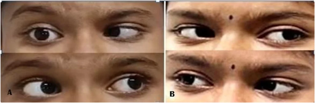
Magnetic resonance imaging revealed the severity of the condition. The child had right-sided otomastoiditis, skull-base osteomyelitis, and erosion of the petrous apex. At the same time, a retropharyngeal abscess measuring approximately 3.1 × 2.2 × 1.4 cm had developed. The collection extended from the petrous apex region toward the nasopharynx. The usual ear findings were not prominent: there was no persistent purulent discharge, and the child was afebrile. Such subtle local signs can mask a deep infection. Therefore, new-onset double vision, retro-orbital pain, or cranial nerve palsy following an ear infection requires thorough clinical evaluation and appropriate imaging.
The doctors surgically drained the abscess the same day, removing approximately 10 mL of pus. They then performed a myringotomy and inserted a ventilation tube to improve middle-ear aeration. Treatment was continued with prolonged antibiotic therapy because of the skull-base osteomyelitis and deep infection. The sixth cranial nerve palsy resolved on the first postoperative day, followed by the resolution of double vision, neck stiffness, and difficulty swallowing. Follow-up magnetic resonance imaging four weeks later showed marked reduction in the inflammatory changes and complete resolution of the abscess.
This case serves as a reminder that an almost normal ear examination does not rule out a serious complication. In children, neurological symptoms may sometimes be the first sign that an infection has spread much deeper than initially suspected.
