

First reported paediatric case of a rare cardiac tumour discovered in a 12-year-old girl
A 12-year-old girl visits a doctor with seemingly common symptoms such as cough, abdominal pain, and dizziness. However, behind these symptoms was something far more serious, a rare primary tumor of the heart.
The case was described in a 2026 case report titled First Paediatric Case of Cardiac Composite Hemangioendothelioma by radiologist Shrinivas Yuvan Shanmuga Sundaram.
The first paediatric case of a rare vascular tumour in an extremely rare location
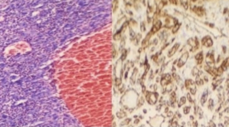
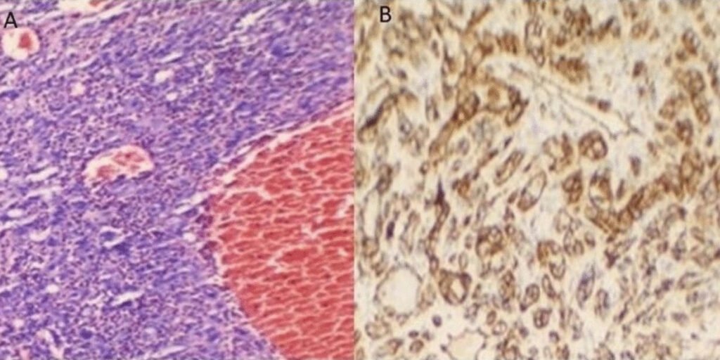
Heart tumors in children are extremely rare, and most of them are not malignant. Using ultrasound, doctors detected a mass in the right atrium, one of the chambers of the heart. At first, an aggressive form of cancer such as angiosarcoma or rhabdomyosarcoma was suspected. However, the final answer came only after examining the tissue under a microscope. The tumor was identified as a composite hemangioendothelioma, an extremely rare vascular tumor, particularly unusual in the heart and exceptionally rare in children.
“Primary cardiac tumours in children are exceptionally rare. Majority are benign, with only about 10% being malignant; rhabdomyosarcoma is reported to be the commonest variety. We present the first paediatric case of a rare vascular tumour in an extremely rare location (right atrium) hitherto unreported in literature.” Shrinivas Yuvan Shanmuga Sundaram et al., case report (2026)
“We utilized a multimodality imaging approach. This included echocardiography, contrast-enhanced computed tomography, and cardiac MRI to diagnose this rare tumor. The findings were confirmed via histopathology and immunohistochemistry of the surgically resected tumor. Additionally, eight weeks post-surgery, the patient underwent an 18F-FDG PET-CT, which is a molecular and functional imaging modality to check for residual tumor and for staging purposes,” explained Dr. Shrinivas Yuvan Shanmuga Sundaram.
Unlike aggressive tumors, this type usually grows more slowly and is less likely to spread to other parts of the body, which means a better prognosis. However, it can recur after treatment, so careful follow-up is necessary. The girl underwent surgery to remove the tumor, after which she began chemotherapy. So far, there are no signs that the disease has spread.
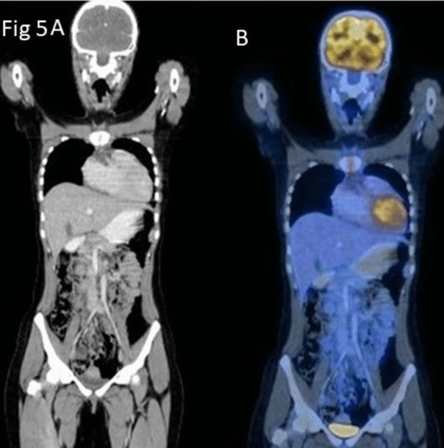
This is a reminder that even when symptoms seem harmless can sometimes point to rare and complex conditions. That is why the combination of modern imaging and precise laboratory analysis remains crucial for making an accurate diagnosis and ensuring successful treatment. “This case highlights how these complementary modalities mutually support the diagnosis and follow-up of this one-of-a-kind cardiac tumor that is the first of its kind in children,” the authors concluded.
Image: Shrinivas Yuvan Shanmuga Sundaram et al., case report (2026)
